przez doddy » 07 Lis 2012, 16:35
Doszły wyniki histopatologiczne Avatara:
Cytological examination 1)
Large nasal lesion (with associated bone lysis) and with enlarged left
mandibular lymph node.
DESCRIPTION:
Three preparations are examined, which display moderate to marked numbers of
nucleated cells and erythrocytes on an often magenta and finely granular
background (mucinous). In the preparations, there is a moderate to marked
amount of basophilic granular material in aggregates (likely necrotic debris).
Most nucleated cells present are macrophages evident singly and in aggregates,
often displaying marked and fine cytoplasmic vacuolation as well as
occasional leukophagia, intracytoplasmic basophilic granular material (cell
debris) as well as occasional erythrophagia, and intracytoplasmic basophilic
and yellow granular material (hemosiderin and hematoidin). Also present
are moderate numbers of variably preserved neutrophils and occasional small
lymphocytes as well as low to moderate numbers of oval to spindeloid and
mild to moderately atypical cells present singly and in aggregates. These
cells display moderate anisocytosis and have a moderate amount of basophilic
cytoplasm with moderate nuclear to cytoplasmic ratios and oval nuclei
displaying mild to moderate anisokaryosis and containing granular chromatin
with small nucleoli (suspected reactive macrophages and fibroblasts - cannot
exclude a neoplastic cell population). Microorganisms are not clearly evident.
INTERPRETATION:
Marked macrophagic and neutrophilic inflammation, likely with necrosis.
Mildly atypical, oval to spindeloid cells evident - cannot exclude neoplasia.
COMMENT:
Necrosis is likely represented, and the inflammatory respose is likely at
least partly due to this process. Potential underlying differentials for
findings would include sepsis (bacterial, fungal) or neoplastic disease.
Microorganisms are not clearly evident, although certain bacteria and
fungi do not stain well with Romanowsky stains.
We will attempt to de-stain the preparations and re-stain with other stains,
which can highlight microorganisms. If we are successful, and there is
additional data, then an addendum will follow.
In light of findings, sampling for running bacterial and fungal culture and
sensitivity is a serious consideration.
The low numbers of mildly atypical oval to spindeloid cells are suspected to
be moderately reactive macrophages, fibroblasts, and possibly osteoblasts in
light of radiographic findings. I cannot completely exclude a neoplastic
process, and biopsy of the lesion remains a consideration for histopathologic
assessment.
ADDENDUM
Two preparations were destained and stained with PAS and an acid-fast stain.
Microorganisms were not encountered in these preparations.
Ponieważ diagnoza jest długa, po angielsku to opiszę w lekkim skrócie.
Avatar miał pobrane 3 próbki w biopsji cienkoigłowej. W próbkach tych stwierdzono przewlekłe stany zapalne, komórki różnego typu także atypowe, dużo zmian martwicowych, w tym kostnych. Badanie wskazuje, że być może być grzybica, a próbki poszły do dalszych badań (wyniki za kolejny miesiąc). Nie wykluczono, nie potwierdzono nowotworu.
Ponieważ jednak stan Avatara zaczyna być coraz poważniejszy, Avatar się dusi, a wycieki krwi z nosa pojawiają się coraz częściej po konsultacjach z 3 lekarzami weterynarii wdrażamy leczenie grzybicy. Obawiamy się, że narośl powoli zatyka przewody nosowe stąd duszności.

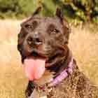
13.10.2011 o 4:45 świat się zatrzymał... Mamba [*]